Let’s Chat About Osteoporosis

On World Osteoporosis Awareness Day this year, our community came together to connect & learn about osteoporosis, and enjoy a delicious calcium-rich cheese platter sponsored by Woombye Cheese Company. Also thank you, IGA Forest Glen! This gathering marked the launch of our “Let’s Chat About Osteoporosis” support and information group! We’re excited to announce that from now on, we will run this support group for our members once a month. It’s a space where we can share experiences, discuss research-based information, and support one another on the journey to better bone health. Here are some important facts about osteoporosis: Osteoporosis weakens bones, making them more brittle and prone to fractures. It usually develops gradually and often shows no symptoms until a fracture occurs. 1 in 3 women and 1 in 5 men over 50 will experience a fracture in their lifetime. Only 20% of patients with osteoporotic fractures are ever diagnosed or treated for the disease. Fun fact: We had so much cheese that our Monday classes got to enjoy some too – all while balancing on a Bosu ball! We look forward to welcoming more of our members to our future group meetups!
World Osteoporosis Day

We’ll be acknowledging World Osteoporosis Day this year by coming together to raise awareness and build a community for those who need support.
Unmasking the Silent Disease: Osteoporosis Risk Factors

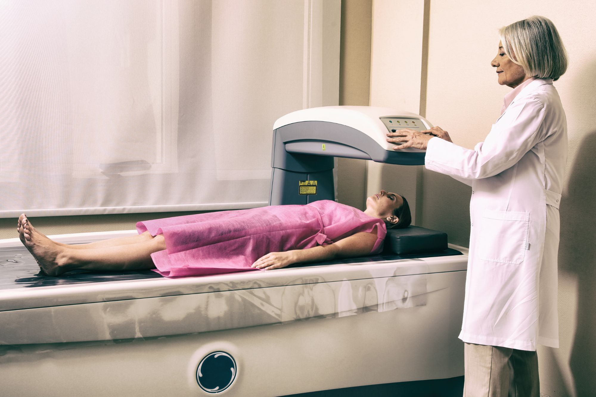
Am I in the risk group for osteoporosis? Osteoporosis is often referred to as a “silent disease” because it typically doesn’t cause noticeable symptoms until a fracture occurs. However, there are certain signs and risk factors associated with osteoporosis that individuals should be aware of. Risk factors for developing osteoporosis Age & gender Family history & genetics Low BMD Hormonal imbalances Nutritional deficiencies & lifestyle choices Lack of exercise Medical conditions/medications Body structure & Low body weight Don’t wait to check until it’s too late Osteoporosis is often not discovered until a bone fracture occurs! The most common symptom of osteoporosis is bone fractures, which can occur with minimal trauma or even from everyday activities like bending, lifting, or falling from standing height. Common fracture sites include the hip, spine (vertebrae), wrist, and shoulder. If you have sustained a low traumatic fracture, but haven’t received the diagnosis of osteoporosis, please read our blog post on ‘diagnosing osteoporosis’, and seek medical assistance. What are the biggest risk factors for Osteoporosis? Risk factors for osteoporosis: Age & gender The risk of osteoporosis increases with age, especially in women after menopause and in men over the age of 70. Although the risk to sustain a low traumatic fracture increases substantially already after the age of 50: 1/3 women, and 1/5 men over the age of 50 will sustain a low traumatic fracture. Women are at a higher risk of developing osteoporosis than men. The hormonal changes during menopause, such as a decrease in Estrogen production, can accelerate bone loss. Men typically do not experience the same rapid loss of bone mass as women do after menopause. Although, by around the age of 70, both men and women start losing bone mass at a similar rate. Risk factors for osteoporosis: Family history & genetics Having a family history of osteoporosis or fractures increases the likelihood of developing the condition. Risk factors for osteoporosis: Low Bone Mass Density (BMD) A lower-than-normal BMD, which can be determined through a bone density test, is a significant risk factor for osteoporosis. Risk factors for osteoporosis: Hormonal imbalances Low levels of Estrogen in women and low levels of testosterone in men can contribute to the development of osteoporosis. Risk factors for osteoporosis: Nutritional deficiencies & lifestyle choices Inadequate intake of essential nutrients for bone health, such as calcium and vitamin D can increase the risk of osteoporosis. Certain lifestyle choices can increase the risk, including excessive alcohol consumption and smoking. Risk factors for osteoporosis: Lack of exercise Insufficient physical activity, whether during childhood, early adulthood, or later in life, is a risk factor for osteoporosis development. Sedentary behavior and inadequate or inappropriate exercise contribute to the increased vulnerability to the condition. Risk factors for osteoporosis: Medical conditions & medications Certain medical conditions, such as rheumatoid arthritis, celiac disease, Crohn’s disease, and hormonal disorders, can increase the risk of osteoporosis. Long-term use of corticosteroids (glucocorticoids) and some other medications can weaken bones and lead to osteoporosis. Risk factors for osteoporosis: Body structure and low weight Small body frame, low body weight, and being underweight are associated with a higher risk of osteoporosis.It’s important to note that having one or more risk factors doesn’t necessarily mean an individual will develop osteoporosis, and individuals without identifiable risk factors can still be affected. However, understanding these risk factors can help individuals and healthcare professionals assess the likelihood of developing osteoporosis and take appropriate preventive measures.
Diagnosing Osteoporosis: Bone Mineral Density and assessment

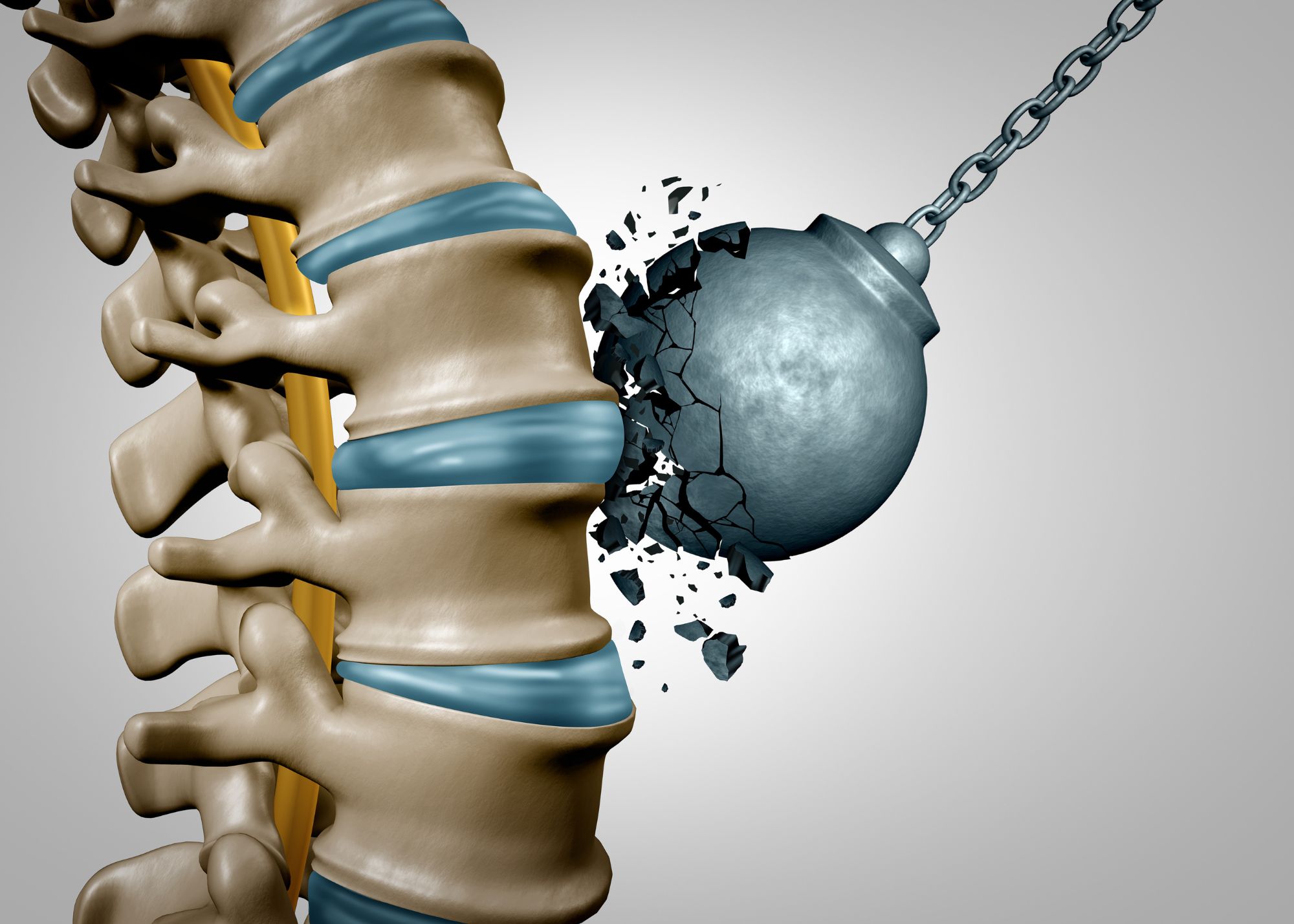
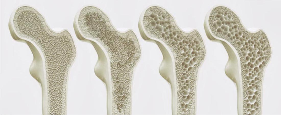
Characteristics of Osteoporosis Osteoporosis is a disease characterised by low bone mass and structural deterioration of bone tissue, leading to increased fragility and susceptibility to fractures. It is estimated that over 200 million people worldwide are living with osteoporosis. Proper diagnosis of osteoporosis is crucial for timely intervention and management of the condition. Bone Mineral Density (BMD) in osteoporosis Bone Mineral Density (BMD) testing is the gold standard for diagnosing osteoporosis. BMD is one determinant of bone strength. This test measures the density of minerals, such as calcium, in the bones. Dual-energy X-ray absorptiometry (DXA) scans are commonly used to assess BMD at the hip, lumbar spine, and wrist. The World Health Organization (WHO) defines osteoporosis as a BMD of 2.5 standard deviations below the mean, typically indicated by a T-score of -2.5 or lower. A T-score between -1.0 and -2.5 indicates osteopenia, a precursor to osteoporosis. While DXA scans are readily available and non-invasive with low radiation exposure, they only provide a two-dimensional measurement of BMD at specific sites, which may not fully represent overall bone health when used alon The natural curve of bone mineral density from childhood to adulthood The natural curve of bone mineral density follows a pattern from childhood to adulthood. During childhood and adolescence, the body typically produces more osteoblasts (cells responsible for bone formation) than osteoclasts (cells responsible for bone breakdown), resulting in a net gain of bone mass and density. However, as we approach our late 20s or early 30s, the rate of bone formation slows down, and the balance shifts towards osteoclasts. This leads to a plateau and possible decline in bone mineral density. There are several factors that will play a role in how quickly our net BMD will decline, which can possibly result in a reduced bone strength and increased risk of fracture. Low traumatic fractures in osteoporosis While BMD testing is valuable, it has limitations when used in isolation to predict fracture risk accurately. To address this, the National Bone Health Alliance has proposed expanding the diagnostic criteria for osteoporosis. They suggest considering low-trauma fractures as diagnostic indicators, even without meeting the traditional BMD criteria. For instance, a low-trauma hip fracture, with or without a BMD test, is considered diagnostic of osteoporosis. The presence of low-trauma clinical vertebral fractures, proximal humerus fractures, or pelvis fractures in individuals with osteopenia may also indicate osteoporosis. Additionally, a low-trauma distal forearm fracture in a person with osteopenia at the lumbar spine or hip may be considered diagnostic in certain cases. Low-trauma fractures, also known as fragility fractures, occur due to minimal force or trauma, such as a fall from standing height or minor impact. These fractures typically occur in individuals with reduced bone strength or underlying conditions like osteoporosis. Common sites for these fractures include the hip, spine, wrist, proximal humerus, and pelvis. In addition to low-trauma fractures, healthcare professionals can utilise the Fracture Risk Assessment Tool (FRAX) to estimate an individual’s 10-year probability of experiencing major osteoporotic fractures or hip fractures. Osteoporosis: Fracture Risk Assessment Tool (FRAX) FRAX incorporates various clinical risk factors, including age, sex, BMI, previous fracture history, parental hip fracture, smoking status, alcohol intake, glucocorticoid use, and the presence of secondary causes of osteoporosis. It also integrates BMD measurements, when available, to provide a more accurate assessment. If an individual’s calculated 10-year probability of hip fracture is equal to or exceeds 3% or the 10-year probability of major osteoporotic fracture is equal to or exceeds 20%, it can support a diagnosis of osteoporosis, even without meeting the traditional BMD T-score criteria. By expanding the diagnostic criteria to include low-trauma fractures and utilising tools like FRAX, a greater number of individuals at elevated fracture risk can be identified. However, it’s important to acknowledge that FRAX has limitations as well. It does not consider certain risk factors such as physical activity, falls, or muscle strength. Moreover, FRAX has primarily been developed for postmenopausal women and men over 50 years old, limiting its applicability to other populations. Testing and Assessment to Diagnose Osteoporosis Diagnosing osteoporosis involves a combination of diagnostic methods, including BMD testing, assessment of low-trauma fractures, and the use of tools like FRAX. While BMD testing remains the gold standard, the inclusion of low-trauma fractures and fracture risk assessment tools helps improve the accuracy of diagnosing osteoporosis and identifying individuals at high risk of fractures. Sunshine Coast Clinic Specialising in Osteoporosis Nordica Health Clinic is an Exercise Physiology clinic on the Sunshine Coast, specialising in helping individuals with Osteoporosis live a quality life with minimal pain. Our Exercise Physiologists use evidence based testing and practices to together with their clients develop an individualised training program, individually and in groups. Contact Nordica Health Clinic in Forest Glen, Sunshine Coast, to find out more how we can assist you on your osteoporosis journey.
Unveiling the Foundation: Exploring the Key Factors of Bone Strength

Bone strength is essential for maintaining a healthy skeletal system and plays a crucial role in supporting our body’s structure, protecting vital organs, and facilitating movement. While bones may appear solid and unyielding, they possess dynamic properties that contribute to their overall strength and integrity. In this post, we will delve into four significant factors that contribute to bone strength: bone mineral density (BMD), bone microarchitecture, bone turnover, and bone quality. Each of these characteristics plays a distinct role in determining the overall strength and resilience of our bones. Bone Mineral Density (BMD) Bone Mineral Density (BMD) is a measure of the amount of minerals (such as calcium) in bone tissue. Higher BMD generally indicates stronger bones. BMD is most commonly measured through a dual-energy X-ray absorptiometry (DXA) scanner at the hip, the lumbar spine, and occasionally the wrist. DXA is accessible, easy to use, and a relatively non-invasive procedure with low radiation exposure. It’s useful for monitoring changes in bone health and assessing fracture risk over time. However, DXA limitations include measuring only two-dimensional BMD at the hip and spine, which may not represent overall bone health accurately. Also, it may not detect changes in bone microarchitecture or quality, which can affect bone strength and fracture risk. Bone microarchitecture Bone microarchitecture refers to the microscopic structure of bone tissue, including the organisation and arrangement of bone cells and the mineralised bone matrix. Good bone microarchitecture typically involves well-organised, dense trabecular (spongy) bone and thick, strong cortical bone. TBS (Trabecular Bone Score) and HR-pQCT (High-Resolution Peripheral Quantitative Computed Tomography) are two different methods for assessing bone microarchitecture. TBS is a software-based technique that uses standard DXA images to provide information on trabecular bone microarchitecture, while HR-pQCT is a specialised imaging technique that creates high-resolution 3D images of both trabecular and cortical bone microarchitecture. TBS is relatively low-cost and non-invasive, while HR-pQCT provides more detailed information on bone microarchitecture. Bone turnover Bone turnover refers to the natural process of bone remodeling, which involves a balance between bone formation and bone resorption, regulated by our bone cells. When this balance is disrupted, such as in the case of excessive bone resorption, bone strength may be compromised. Bone turnover can be assessed by measuring markers of bone formation and resorption. These markers can be measured using blood and urine tests, and they provide information on the rate of bone turnover. Examples of bone formation markers include osteocalcin, P1NP, and BSAP, while examples of bone resorption markers include CTX, NTX, and DPD. However, it’s important to note that these markers can be influenced by various factors such as age, sex, menopausal status, certain medications, and medical conditions. Bone quality Bone quality is a broad term that encompasses several factors, including the composition and organisation of bone tissue, the number and organisation of bone cells, and the overall health and function of bone tissue. Bone quality can be assessed using a variety of methods, including bone biopsy, imaging techniques, and biochemical markers. Imaging techniques such as DXA, HR-pQCT, and TBS can provide indirect measures of bone quality by assessing bone density, microarchitecture, and texture, respectively. Biochemical markers, such as advanced glycation end products (AGEs), can provide information on the quality of the bone matrix. Other factors that can impact bone quality include the composition and organisation of the bone tissue, the presence of microdamage or other defects, and the presence of comorbidities that can affect bone health, such as kidney disease or diabetes. Bone mineral density (BMD) serves as a fundamental measure of bone health, reflecting the mineral content within bone tissue. However, while BMD measurements through DXA scanning provide accessible and non-invasive assessments, they may not capture the full complexity of bone health, such as changes in microarchitecture and quality. A balance between bone formation and resorption is important for maintaining healthy bone turnover, while bone quality depends on several factors related to bone tissue composition and organisation. Higher bone mineral density and well-organised, dense trabecular and cortical bone are all factors influencing bone strength.
Bone Anatomy and Physiology: Understanding the Continuous Process of Bone Remodeling

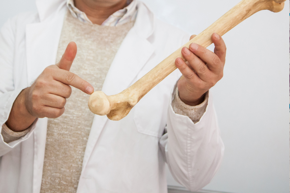
Are you curious about the structure and function of our bones? Understanding the anatomy and physiology of healthy bone tissue is essential to grasp the pathophysiology of osteoporosis. Let’s take a closer look at this fascinating topic! Bone Anatomy – the structure of bone Our bones are made up of cells, fibers, and a mineralised matrix that gives them strength and rigidity. The bone cells include osteoblasts, osteocytes, and osteoclasts, which respectively build, engineer, and recycle bone tissue. The bone matrix is composed of collagen fibers and mineral crystals, such as calcium and phosphorus, that provide the bone with its unique properties. The periosteum is a thin layer of connective tissue that covers the outer surface of bones and contains blood vessels and nerves. The medullary cavity is a hollow space in the center of long bones that contains bone marrow, which produces blood cells. Bone Physiology – the function of bone Bones grow through ossification, a process that involves the formation of new bone tissue by osteoblasts. This process occurs throughout childhood and adolescence and is regulated by various hormones, such as growth hormone and estrogen. Bone remodeling is a continuous process of breaking down and rebuilding bone tissue, essential for maintaining bone strength. This process is regulated by hormones such as parathyroid hormone and calcitonin. Bones play a crucial role in maintaining calcium homeostasis in the body. When blood calcium levels are low, calcium is released from bones through the action of osteoclasts. When blood calcium levels are high, calcium is deposited back into bones through the action of osteoblasts. Bones can repair themselves after injury through a process called bone healing, which involves the formation of new bone tissue by osteoblasts. Mechanotransduction Interestingly, bones also have the ability to adapt to mechanical stress, through a process called mechanotransduction. This is where cells convert mechanical signals produced by external loading into biochemical signals that regulate cellular activity. This adaptation is crucial to regulating bone remodeling in response to stress and maintaining bone strength. Overall, understanding the anatomy and physiology of bone tissue provides a foundation for understanding the pathophysiology of bone diseases such as osteoporosis. The complex balance between bone formation and resorption is essential to maintaining bone strength and function. So, take good care of your bones, and don’t forget to learn some more on how you can optimise your cellular mechanotransduction!
Osteoporosis and Successful Aging: Psychosocial Interventions for Improved Quality of Life

Building Strength and Resilience with Osteoporosis Despite advancements in the diagnosis and treatment of osteoporosis, there is still much work to be done. Current protocols predominantly focus on pharmacologic intervention, overlooking the holistic needs of patients. This narrow approach fails to address the extensive physical, financial, and psychosocial ramifications of the disease, particularly when a low traumatic fracture occurs. The National Institutes of Health Consensus Conference on Osteoporosis has recognized that osteoporosis affects individuals of all ages, genders, and races. The consequences of this disease are far-reaching and can impact not only the individual but also their family and community. This blog post aims to explore the psychological and social implications of osteoporosis in postmenopausal women. Additionally, we propose effective strategies to manage these outcomes and highlight future research directions to improve the overall well-being and quality of life for all individuals affected by osteoporosis with a low traumatic fracture. Osteoporosis is a hidden disease that can have significant sociopsychological implications on individuals, with potential impacts on their quality of life, mental health, and social well-being. Fear of falling, social isolation, depression, anxiety, body image issues, and stigma are some of the common concerns that individuals with osteoporosis may experience. This fear increases if the individual also has experienced a low traumatic fracture. It is therefore important to raise awareness about the psychosocial interventions that could impact health perceptions at the onset of osteoporosis and help people negotiate changes that are associated with osteoporosis and the aging process itself. Fear of falling and fear avoidance One of the most significant concerns for people with osteoporosis is the fear of falling. This fear can lead to decreased physical activity, social isolation, depression, and anxiety. This can further contribute to decreased bone strength and increased risk of falls. Therefore, regular physical activity can help to reduce bone loss and increase bone strength, thereby reducing the risk of fractures. Exercise can also help to improve balance and coordination, and confidence which can reduce the risk of falls, fractures and psychosocial issues. Body image and stigma Women with osteoporosis often report feeling self-conscious about their appearance and perceive themselves as “fragile” or “weak.” This can lead to a negative impact on self-esteem and mental health. A study on the impact of osteoporosis on quality of life found that individuals with osteoporosis reported feeling stigmatised and perceived as “old” or “feeble,” leading to negative feelings about their self-worth and social identity. Support groups can provide emotional and social support for people with osteoporosis, helping them to cope with the challenges of the disease and maintain a positive outlook. Raising awareness about the sociopsychological impact of osteoporosis can help reduce stigma and promote understanding and empathy towards individuals living with this condition. Psychosocial interventions Education plays a crucial role in supporting individuals with osteoporosis, covering a range of topics like disease understanding, prevention, testing, and available treatments. These educational programs, benefiting both diagnosed and undiagnosed individuals, are vital for promoting disease prevention and overall well-being. Through these educational initiatives, individuals can gain knowledge about prevention, rehabilitation, the impact of aging on the body and mind, exercise and nutrition, retirement planning, combating ageism, life transitions, and end-of-life considerations: Learning about osteoporosis Education is one of the most important psychosocial interventions for osteoporosis. Health promotion and disease prevention programs have an important role in successful aging. Educational programs should address all aspects of the disease process such as what osteoporosis is, what are the physiologic implications of the disease, who is at risk, how can osteoporosis be prevented, how to get tested, and what therapies for treatment are available. Programs that focus on providing information to people who have or have not been diagnosed with osteoporosis are extremely important. Knowledge can be powerful in the disease prevention and health promotion process. Promoting a successful aging mindset Successful aging involves low risk of disease and disease-related disability, high mental and physical function, and active engagement in life. Educational programs dealing with successful aging are imperative and should be readily available to the community. Some of the issues that could be addressed include disease prevention and rehabilitation through proper detection and treatment, physiologic and psychological effects of aging, proper exercise and nutrition, retirement living, ageism, life course transitions, and death and dying. Enhancing Quality of Life with Osteoporosis In conclusion, osteoporosis, particularly when recovering from a low traumatic fracture, can be a debilitating disease that affects a person’s ability to carry out daily activities. However, with proper education, exercise, support, and housing modifications, people with osteoporosis can maintain a good quality of life and age successfully. It is important to raise awareness about the psychosocial interventions that could impact health perceptions at the onset of osteoporosis and help people negotiate changes that are associated with osteoporosis and the aging process itself. By focusing on the psychosocial implications of osteoporosis, healthcare providers can join us in providing appropriate support and resources to address these concerns, ultimately improving the quality of life of those living with this condition. Sunshine Coast Clinic Specialising in Osteoporosis Nordica Health Clinic is an Exercise Physiology clinic on the Sunshine Coast, specialising in helping individuals with Osteoporosis live a quality life with minimal pain. Our Exercise Physiologists use evidence based testing and practices to together with their clients develop an individualised training program, individually and in groups. Contact Nordica Health Clinic in Forest Glen, Sunshine Coast, to find out more how we can assist you on your osteoporosis journey.
May is Osteoporosis Awareness Month!

What is the Osteoporosis Awareness Month? Throughout the month of May, we invite you to join us on a bone health journey. Together with our team and members, we will provide information and resources to increase awareness of osteoporosis, a common condition that weakens bones and makes them more prone to fractures. Our aim is to empower you with the knowledge and tools you need to make small but impactful lifestyle changes to improve or maintain your bone health and longevity. Maintaining good bone health is essential to our overall well-being and quality of life as we age. Our bones provide the structure for our other structures and systems, which is why it’s important to take proactive steps early on to keep them healthy and strong for as long as possible. What can I do to improve my osteoporosis? Maintaining good bone health is essential to our overall well-being and quality of life as we age. Our bones provide the structure for our other structures and systems, which is why it’s important to take proactive steps early on to keep them healthy and strong for as long as possible. At Nordica Health we complete a health screen and a thorough assessment will help tailor the exercise plan to your individual needs and preferences. Your starting point and maintenance phase will depend on your risk factors and individuality. Taking action to improve your bone health can reduce the risk of low traumatic fracture and improve your overall quality of life.
Osteoporosis and Osteopenia

What is osteoporosis? Osteoporosis is a condition that weakens the bones and increases the risk of low traumatic fracture. The gold standard for diagnosing osteoporosis is the DXA scan, which measures Bone Mineral Density and provides a classification system to identify osteoporosis or osteopenia. Common body sites that are measured include the neck of femur, vertebrae of the lumbar spine, and occasionally the wrist. Low traumatic fractures are a significant risk worldwide, with 1 in 4 women and 1 in 5 men over 50 years old expected to sustain a fracture at some point in their lives. The impact of a fracture on an individual can result in fear avoidance of certain activities, loss of confidence, reduced mobility, strength, and functional capacity, a reduced quality of life, and depression or anxiety. How is osteoporosis assessed and diagnosed? Osteoporosis is a silent disease, and many people are unaware they have it until they sustain a fracture. If you have one or more risk factors, it is a good idea to discuss this with your GP and get a bone health assessment. Early diagnosis can help you take actions to improve your bone health, such as optimizing your diet, spending time in the sun, quitting smoking, reducing alcohol consumption, and commencing and maintaining a consistent osteogenic exercise routine. Osteoporosis can be diagnosed through three methods, which require confirmation that there is no other condition that appears to be osteoporosis but is not. The first method is a bone mineral density test that produces a T-score, with a score of -2.5 or below indicating osteoporosis. The second method is through a fracture, especially of the spine or hip, which suggests weak bones and the need for further evaluation. The third method involves using the FRAX fracture risk calculator, which assesses the chances of having different types of fractures in the next 10 years based on bone density, age, sex, height, weight, and answers to 7 questions. If the probability of a major osteoporotic fracture or hip fracture is above certain thresholds, a diagnosis of osteoporosis can be made. I have been diagnosed with osteoporosis, what do I do now? Exercise is one of the most effective non-pharmacological lifestyle changes that can influence bone strength and reduce the risk of fracture and falls, increase functional strength, improve confidence, reduce anxiety and depression, and improve overall quality of life. The Australian guidelines for physical activity recommend a minimum of 150 minutes per week of moderate-intensity aerobic exercise and at least two resistance training sessions per week to maintain a healthy life. However, exercise recommendations for osteoporosis focus on progressive resistance training, progressive impact training, and balance type activities. An Exercise Physiologist can help osteoporosis To optimize your bone health exercise plan, seek out an Accredited Exercise Physiologist or Physiotherapist who specializes in exercise for osteoporosis. At Nordica Health we complete a health screen and a thorough assessment will help tailor the exercise plan to your individual needs and preferences. Your starting point and maintenance phase will depend on your risk factors and individuality. Taking action to improve your bone health can reduce the risk of low traumatic fracture and improve your overall quality of life.
Osteoporosis awareness day

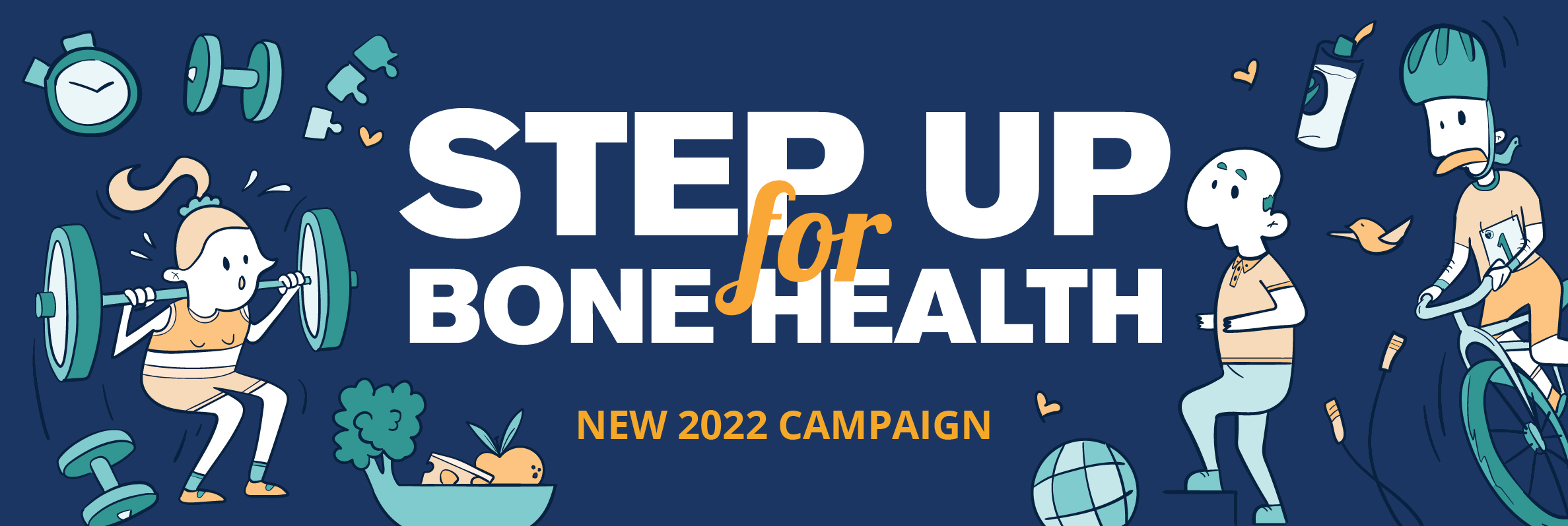
Step Up For Bone Health Today, Thursday the 20th of October is world osteoporosis awareness day, and we joined the campaign Step Up For Bone Health. Greta and I went to Cotton Tree Park to run our event “moving for healthy bones” to increase the awareness of what osteoporosis is, to identify risk factors, and to promote healthy lifestyle choices to improve bone health. Jessica interviewed on 109.4 (SunshineFM) for osteoporosis awareness day Exercise Physiologists Greta and Jessica talked about osteoporosis, and ran a movement session “Moving For Healthy Bones” on Osteoporosis Awareness Day. What is osteoporosis and osteopenia? Osteoporosis is a condition that weakens the bones, and increases the risk of low traumatic fracture. According to the World health organization, the gold standard to diagnose osteoporosis is through the DXA scan, which measures Bone Mineral Density, and provides a classification system to identify osteoporosis, or osteopenia. The common body sites that are measured are the neck of femur (hip) of non-dominant side, the vertabraes of the lumbar spine, and occasionally the wrist. Low traumatic fractures Worldwide; 1 in 4 women, and 1 in 5 men over 50 years old, will sustain a low traumatic fracture at some point in their life. Common fracture sites include; the vertebrae, the neck of the Femur (hips), wrists, ankles, toes, ribs. The impact of a fracture on somebody will depend on various factors, but can result in fear avoidance of certain activities, loss of confidence, reduced mobility, strength, and functional capacity; a reduced quality of life, depression and/or anxiety. Are you at risk? Risk factors for osteoporosis The majority of people who have osteoporosis will be unaware that they have it, until they sustain a fracture. This is why it is called a ‘silent disease’. If you have one or more risk factors, it might be a good idea to discuss this with your GP, and to get a bone health assessment. You can do so much more to increase your bone health by getting diagnosed early; before sustaining a fracture. Actions to improve your bone health includes: optimizing your diet (how, depend on your needs), spend 10-15 min in the sun per day, cease smoking, reduce alcohol consumption, and commence + maintain a consistent osteogenic exercise routine. Exercise prescription for osteoporosis: improve your bone health Exercise is one of the most effective non-pharmacological lifestyle change you can do that can not only substantially influence your bone strength, but can also reduce the risk of fracture and risk of falls, increase functional strength, improve your confidence, improve anxiety and depression, and improve your overall quality of life. The Australian guidelines of physical activity to maintain a healthy life include 1. Aerobic type exercise, a minimum of 150 min per week, at moderate exercise intensity, and 2. Resistance training, at least two times per week. However, if we are solely looking at exercise recommendations for osteoporosis, the guidelines are somewhat different. When implementing an exercise routine to assist in managing your osteopenia, osteoporosis, or preventing a reduction in bone strength, you can generally split them into three main categories. Progressive Resistance Training Progressive Impact Training Balance type activities Your starting point, and “maintenance point” (when you reach the maintenance phase), will be dependent on your risk factors, and your individuality. Get expert advise for your bone health exercise plan To optimize your bone health exercise plan, it would be a good idea to go through a health screen and assessment first. The exercise plan can then be tailored to suit your level of risk, preference and needs. We therefore recommend you seek out an Accredited Exercise Physiologist or Physiotherapist, who specializes in exercise for osteoporosis, before commencing an exercise plan to improve your bone health. Take the quiz Osteoporosis, and the risk of low traumatic fracture affects millions of people in the world, and you could be one of them. Take the quiz on worldosteoporosisday.org to identify any risk factors. If you are at risk, please discuss with your GP, and start implementing choices that your bones will thank you for. Get those bones moving; give them the specific exercise prescription they need.